Human Eye Structure and Function PDF
Hello Aspirants,
The human eye is a complex organ responsible for vision. Here’s an overview of its structure and function:
Structure:
Cornea: The transparent, dome-shaped outermost layer that covers the front of the eye. It helps focus light onto the retina.
Iris: The colored part of the eye that surrounds the pupil. The iris controls the size of the pupil and regulates the amount of light entering the eye.
Pupil: The black circular opening in the center of the iris. It changes size based on the amount of light present, allowing more or less light to enter the eye.
Lens: Located behind the iris, the lens focuses light onto the retina. It can change shape to adjust the eye’s focus, a process known as accommodation.
Retina: The innermost layer at the back of the eye that contains specialized cells called photoreceptors. These photoreceptors, known as rods and cones, convert light into electrical signals that are sent to the brain for visual processing.
Optic Nerve: The bundle of nerve fibers that carries visual information from the retina to the brain, allowing us to perceive and interpret what we see.
Vitreous Humor: A gel-like substance that fills the center of the eye, providing shape and support to the eyeball.
Sclera: The tough, white outer layer of the eye that maintains the eye’s shape and protects the internal structures.
Function:
Light Refraction: The cornea and lens work together to refract (bend) incoming light and focus it onto the retina. This process helps create a clear and focused image.
Accommodation: The lens adjusts its shape to focus on objects at different distances. When viewing nearby objects, the lens becomes thicker, and for distant objects, it becomes thinner.
Photoreception: The retina contains millions of rods and cones. Rods are more sensitive to light and are responsible for vision in low-light conditions, while cones are responsible for color vision and visual acuity in brighter conditions.
Signal Transmission: When light strikes the photoreceptors, they generate electrical signals. These signals are then transmitted to the brain through the optic nerve, where they are processed and interpreted to create visual perception.
Pupillary Reflex: The iris controls the size of the pupil, regulating the amount of light entering the eye. In response to varying light conditions, the pupil constricts (becomes smaller) in bright light and dilates (becomes larger) in dim light.
Binocular Vision: Humans have two eyes positioned slightly apart, allowing for binocular vision. This means that both eyes work together to provide depth perception and a three-dimensional view of the world.
Color Vision: Cones in the retina contain pigments that respond to different wavelengths of light, enabling us to perceive colors. There are three types of cones that are sensitive to different ranges of light wavelengths, allowing us to see a wide spectrum of colors.
The human eye is a remarkable organ that plays a crucial role in our ability to see and perceive the world around us.
- General Science ( सामान्य विज्ञान )Notes in Hindi PDF
- 500 General Science Question Answer In Hindi PDF
- General Science Book PDF For Competitive Exams PDF
- Chemistry Handwritten Notes In Hindi PDF Download
- Physics Handwritten Notes In Hindi PDF
- Biology Handwritten Notes PDF in Hindi Free Download
- General Science Handwritten Notes PDF in Hindi
- General Science book for Competitive Exams PDF Download
- Drishti IAS science and technology book pdf
Most Important Human Eye Structure Question Answer
About Human Eye:
The human eye is one of five sense organs which is t h e m o s t valuable and sensitive.
It helps us to see the wonderful surrondings and the colours around us.
The eyeball is approximately spherical in shape with a diameter of about 2.3 cm to 2.5 cm.
Refraction of the light rays entering the eye mostly occurs at the outer surface of the cornea.
The crystalline convex lens in eye merely provides the finer adjustment of focal length required to focus.
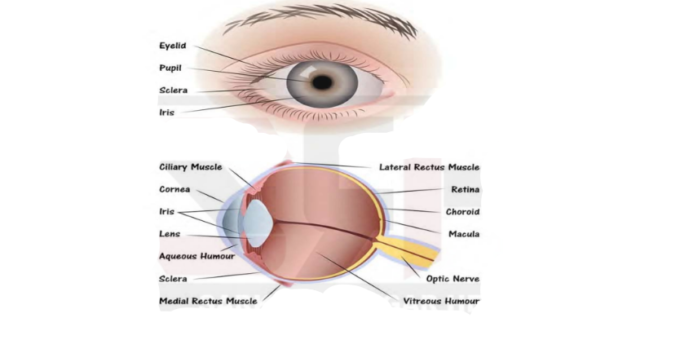
1 Parts Of human Eye:
1. Cornea: The transparent spherical membrane covering the front of the eye.
2. Iris: The coloured diaphragm between the cornea and lens.
3. Pupil: The small hole in the iris.
4. Eye lens: It is a transparent lens made of jelly like material.
5. Ciliary muscles: These muscles hold the lens in position.
6. Retina: The back surface of the eye.
7. Blind spot: The point at which the optic nerve leaves the eye. An image formed at this point is not sent to the brain.
8. Aqueous humour: A clear liquid region between the cornea and the lens.
9. Vitreous humour: The space between eye lens and retina is filled with another liquid called vitreous humour.
In the eye, the image is formed on the retina by successive refractions at the cornea, the aqueous humour, the lens and the vitreous humour.
Electrical signals then travel along the optic nerve to the brain to be interpreted.
In good light, the yellow spot is most sensitive to detail and the image is automatically formed there.
Accommodation:
The ability to focus both near and distant objects, by adjusting its focal length, is called the accommodation Power of the eye
It is the ability of the ciliary muscles to change the focal length of the eye lens, called accommodation. Defects of the Eye:
Although the eye is one of the most remarkable organs in the body, it can have several abnormalities, which can often be corrected with eyeglasses, contact lenses, or surgery.
The various defects that suffer the eye are
o Hypermetropia or long sightedness,
o Myopia or shortsightedness
o Astigmatism, and
o Presbyopia.
Hypermetropia or Long Sightedness:
A person suffering from this defect can see distant objects clearly but cannot see nearby objects clearly.
In this defect, the near point lies farther away than 25 cm.
Hypermetropia (far or long sightedness — the image of nearby objects is focused beyond the retina) is
corrected by using a convex Lens of suitable power.
The eye loses its power of accommodation at old age.
Causes of Hypermetropia:
Either the hyperopic eyeball is too short or the ciliary muscle is unable to change the shape of the lens enough to properly focus the image i.e. the focal length of the eye lens increases.
Myopia or Short Sightedness or Near Sightedness:
A person suffering from myopia or short sightedness can see nearby objects clearly but cannot see the far away objects clearly.
Myopia (short sightedness — the image of distant objects is focused before the retina) is corrected by using a concave lens of suitable power.
Causes of Myopia:
Either the eyeball is longer than normal or
The maximum focal length (due to excessive curvature of the cornea) of the lens is insufficient to produce a clearly formed image on the retina.
Astigmatism:
A person may also have an eye defect known as astigmatism, in which light from a point-source produces a line image on the retina.
A person suffering from this defect cannot see in all directions equally well i.e., he cannot see the vertical and horizontal lines
simultaneously.
This condition arises either when the cornea or the crystalline lens or both are not perfectly spherical.
Astigmatism can be corrected with lenses having different curvatures in two mutually perpendicular directions i.e., cylindrical lens.
When a person suffers from both, the myopia as well as Hypermetropia, his spectacles for correction have bifocal lenses.
The upper half is a concave lens for distant vision and lower half is a convex lens for reading.
Presbyopia:
Presbyopia is that defect of human eye, due to which an old person cannot read and write comfortably.
That is why Presbyopia is also called old sight.
To correct Presbyopia, an old person has to use spectacles with a convex lens of suitable focal length, or power as explained already.
The cause of Hypermetropia is a decrease in length of eyeball or increase In focal length of eye lens.
But the cause of Presbyopia is an increase in focal length of eye lens only.
The eyeball, in Presbyopia, has normal length. the vision of the eye decreases, leading sometimes to total loss of vision.
The problem is overcome by cataract surgery i.e., removal of the eye lens, and its replacement by a lens of suitable focal length.
We need two eyes because a human being has a horizontal field
of view of about 150° with one eye and of about 180° with two eyes.
Thus, two eyes provide us wider horizontal field of view.
With one eye, the world looks flat, i.e., two dimensional only.
With two eyes, the view is three dimensional, i.e., dimension of depth is added to our view.
As our two eyes are separated by a few centimeters, each eye observes a slightly different image.
Our brain combines the two views into one and we get to know how close or far away the things seen are.
D o n a t i o n o f t h e H u m a n E y e:
By donating our eyes after we die, one pair of our eyes can give vision to two corneal blind people.
Eye donors may belong to any sex or any age group.
People suffering from diabetes, hypertension, asthma or any other non- communicable diseases can also donate
their eyes.
People who have been using spectacles or those operated for cataract can also donate eyes.
N e a r P o i n t:
It is the smallest distance, at which the eye can see objects clearly without strain, is called the near point of the eye or the least distance of distinct vision.
For a young adult with normal vision, it is about 25 cm from the eye.
F a r P o i n t:
The farthest point up to which a short-sighted eye can see clearly is called the far point of the eye.
For a normal eye, the far point is infinity.
P e r s i s t e n c e o f V i s i o n o f T h e E y e :
The image of an object persists on the retina for 1/16 second, even after the removal of the object.
The sequence of still pictures taken by a movie camera is projected on a screen at a rate of about 24 images or more per second.
The successive impressions of images on the screen appear to merge smoothly into one another to give us the feeling of moving images.
C o l o u r B l i n d n e s s:
The maximun numbers of light sensitive cells contained in the retina of the eye are of two types: Rod cells and Cone cells.
Rod shaped cells which respond to brightness or intensity of light and Cone shaped cells, which respond to colour of the light. Therefore, cone shaped cells enable us to distinguish between different colours.
When a person cannot distinguish between different colours, he is said to be colour blind though his vision may otherwise be normal.
Colour blindness is a genetic disorder, which occurs by inheritance.
So far, there is no cure for colour blindness.
L e a s t D i s t a n c e o f D i s t i n c t V i s i o n:
The minimum distance up to which an eye can see clearly is called the legist distance of distinct vision.
It is normally denoted by D.
The least distance of distinct vision is equal to the distance between the eye and its near point.
For a normal human eye, this distance is around 25 cm.
The distance between far point and near point of the eye is called range of vision of the eye.
S p e c t r u m:
When white light passes through a prism, the violet light bends most, and the red light bends the least.
Dispersion of light is the phenomenon of splitting of white light into its constituent seven colours on passing through a glass prism.
The band of seven colours so obtained is called visible spectrum.
The seven colours of white light are violet, indigo, blue, green, yellow, orange and red. It is remembered by the acronym VIBGYOR.
Isaac Newton was the first to use a prism to obtain a spectrum of sunlight.
Spectrum is the band of distinct colours we obtain when white light is split by a prism.
C a u s e o f D i s p e r s i o n:
Every colour has its own characteristic wavelength/frequency.
Different colours move with same speed in air/vacuum.
But their speeds in refracting media like glass are different.
Therefore, refractive index of the medium for different colours is different.
As a result, different colours undergo different deviations on passing through the prism.
Hence, different colours emerge from the prism along different directions.
The speed of light in vacuum is same for all wavelengths, but the speed in a material substance is different for different wavelengths.
In any medium other than air/vacuum red light travels the fastest and violet light travels the slowest.
E l e c t r o m a g n e t i c R a d i a t i o n:
The most familiar form of electromagnetic radiation may be defined as that part of the spectrum that the human eye can detect.
Light is produced by the rearrangement of electrons in atoms and molecules.
The various wavelengths of visible light are classified with colours ranging from violet (λ = 4 x 10⁻⁷ m) to red (λ = 7 x 10⁻⁷ m).
The eye’s sensitivity is a function of wavelength, the sensitivity being
a maximum at a wavelength of about λ = 5.6 x 10⁻⁷ m (yellow-green).
When we pass white light through two identical prisms held side by side with their refracting edges in opposite directions; the first prism disperses
white light into seven colours and the second prism recombines the seven colours into white light. Thus, light emerging from 2nd prism is white.
A rainbow is formed due to dispersion of light by tiny droplets of water which act as prisms.
Atmospheric refraction is the cause of twinkling of stars, advance sunrise and delayed sunset.
Scattering of light causes the blue colour of sky and the reddening of the Sun at sunrise and sunset. Some Important previous year
Questions:
Q-1. Which cell disorder in our body is responsible in colour blindness?
(A) WBC (B) Cone cell
(C) Rod Cell (D) Neuron
Q-2. The least distance of distinct Vision is
(A) 35 cm (B) 25 cm
(C) 45 cm (D) 15 cm
Q-3. Sensitivity of human eye is maximum in the
(A) Violet region (B) Green region
(C) Blue region (D) Red region
Q-4. The part of the eye having largest refractive index is
(A) Cornea (B) Aqueous humor
(C) Lens (D) Virtuous humor
Q-5. Short-sight in human eye can be corrected by using proper
(A) Convex lens (B) Concave lens
(C) Cylindrical lens (D) Bifocal lens
Q-6. Colour blindness defect can be corrected by using the lens–
(A) Concave Lens (B) Convex Lens
(C) Cylindrical lens (D) None of these
More Related PDF Download
Maths Topicwise Free PDF >Click Here To Download |
English Topicwise Free PDF >Click Here To Download |
GK/GS/GA Topicwise Free PDF >Click Here To Download |
Reasoning Topicwise Free PDF >Click Here To Download |
Indian Polity Free PDF >Click Here To Download |
History Free PDF > Click Here To Download |
Computer Topicwise Short Tricks >Click Here To Download |
EnvironmentTopicwise Free PDF > Click Here To Download |
UPSC Notes >Click Here To Download |
SSC Notes Download > Click Here To Download |
Topic Related PDF Download
pdfdownload.in will bring you new PDFs on Daily Bases, which will be updated in all ways and uploaded on the website, which will prove to be very important for you to prepare for all your upcoming competitive exams.
The above PDF is only provided to you by PDFdownload.in, we are not the creator of the PDF, if you like the PDF or if you have any kind of doubt, suggestion, or question about the same, please send us on your mail. Do not hesitate to contact me. [email protected] or you can send suggestions in the comment box below.
Please Support By Joining Below Groups And Like Our Pages We Will be very thankful to you.
- Facebook Page: https://www.facebook.com/onlyupsc/